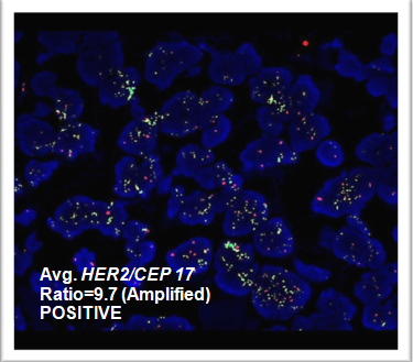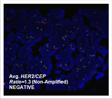
Fluorescence in situ Hybridization (FISH) analysis offers one of the most sensitive, specific reliable and fast strategy for identifying acquired chromosomal changes for enhanced diagnosis and better management of patient. FISH technique has proved to be very useful in many hematological disorders (CML, ALL, AML, Multiple Myeloma), solid tumors (breast cancer, lung cancer, neuroblastoma, burkits lymphoma) and detecting suspected cases of genetic disorders causing birth defects (downs syndrome) both prenatally and postnatal after birth.
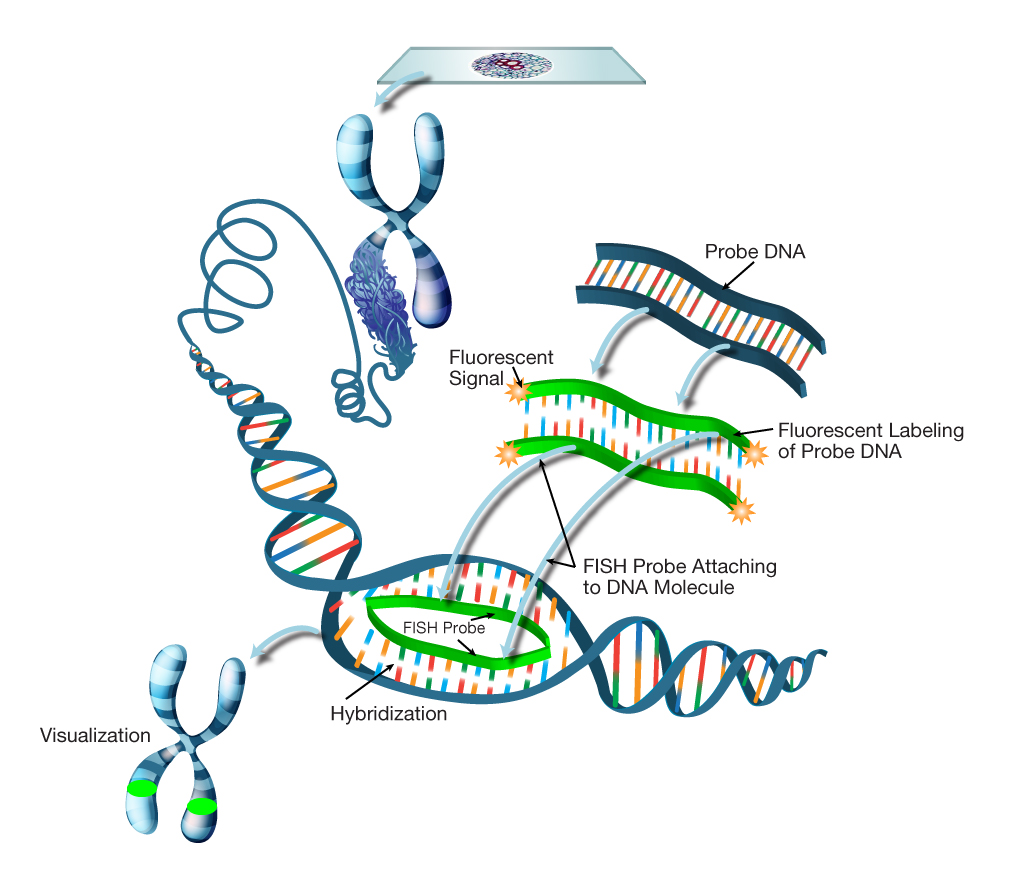
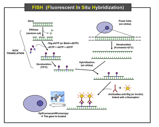
Figure: The schematic diagram of FISH process showing labeling of BAC/PAC clones with fluorescent dyes for the targeted gene followed by procedure to detection on the target sample
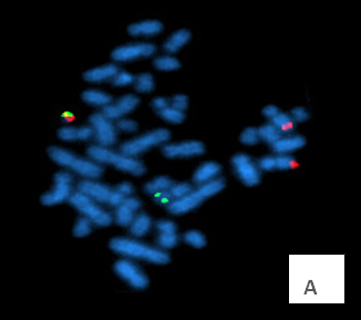
Figure: (A) Metaphase FISH for detection of BCR-ABL gene fusion (B) Interphase cell showing FISH signal with 1 F, 2 R, 1 G signal for BCR-ABL gene in chronic myeloid leukemia (CML) patient.